
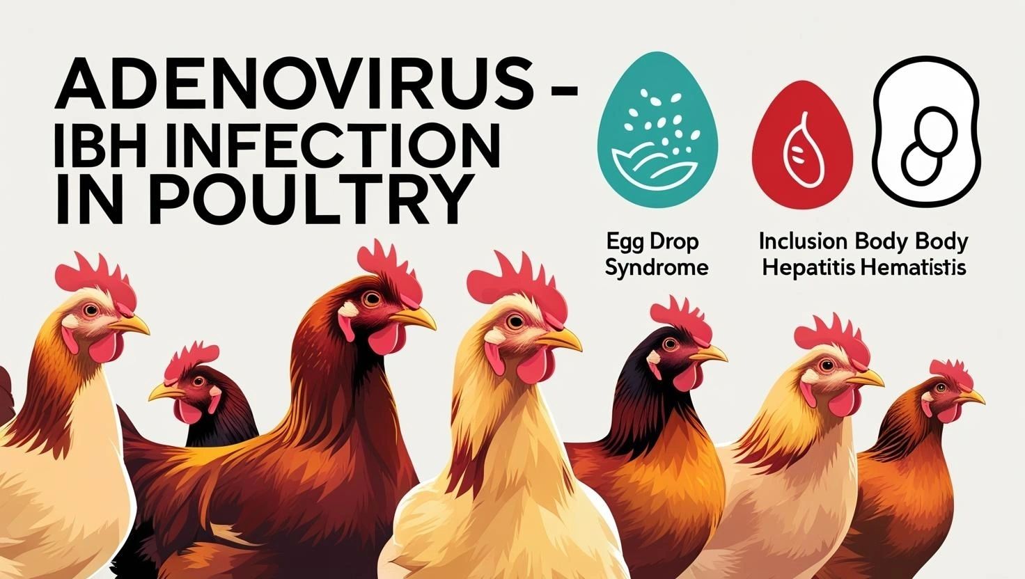
Understanding Avian Adenovirus Infections in Chickens: A Comprehensive Guide
Avian adenovirus infections are a common concern in poultry, particularly affecting chickens worldwide. While many infections can be subclinical or associated with nondescript syndromes, certain adenoviruses are significant primary pathogens causing distinct and economically important diseases. Understanding these infections is crucial for maintaining flock health and productivity.
What are Avian Adenoviruses in Chickens?
Adenoviruses are DNA viruses that replicate and produce characteristic inclusion bodies in the nuclei of infected cells. They are non-enveloped and range in size from 70 to 90 nm. In poultry, adenoviruses are classified into three genera, with those affecting chickens primarily belonging to:
- Aviadenovirus (Group I Avian adenoviruses): This genus includes Fowl adenoviruses (FAdV) with 12 serotypes (FAdV-1 to FAdV-11) assigned to five species (A-E).
- Siadenovirus (Group II Avian adenoviruses): Turkey adenovirus A (TAdV-3), which causes Marble Spleen Disease in pheasants, can also cause Avian Adenovirus Splenomegaly in chickens.
- Atadenovirus (Group III Avian adenoviruses): Duck adenovirus-1 (DAdV-1) causes Egg Drop Syndrome (EDS) in chickens.
How do Chickens Get Infected?
Adenoviruses can spread through both vertical and horizontal transmission.
- Vertical Transmission: Infected breeders can transmit the virus through eggs to their progeny. This can lead to outbreaks in young birds or latent infections that reactivate later, particularly during stress or peak egg production.
- Horizontal Transmission: The virus is shed in feces, making the fecal-oral route a primary mode of spread. Contaminated environments, fomites (such as egg trays and transport vehicles), personnel, and even dust created during cleaning can facilitate spread between farms or within flocks. Some adenoviruses, like FAdV-4, can also spread via aerosol transmission.
Major Adenoviral Diseases in Chickens and Their Lesions
The lesions associated with avian adenovirus infections vary significantly depending on the specific virus or syndrome involved. Here's a breakdown of the key diseases in chickens:
1. Inclusion Body Hepatitis (IBH)
- Definition: IBH is a disease of young chickens characterized by sudden onset, increased mortality, and hepatitis with intranuclear inclusion bodies.
- Causative Agents: Most commonly caused by FAdV-2, FAdV-8 (8a and 8b), and FAdV-11, but also by FAdV-4 (as part of HPS) and FAdV-6.
- Occurrence: Worldwide distribution, typically seen in 2 to 5-week-old broilers, but can affect younger or older birds, layers, and breeders.
- Clinical Signs: Often, the first sign is a sudden marked increase in mortality (up to 30%, but typically lower). Affected birds may be depressed, listless, pale (comb, wattles, facial skin), and show ruffled feathers or huddling. Yellow, mucoid droppings may also be observed.
- Gross Lesions:
- Liver: Consistently affected, appearing swollen, enlarged, pale pink to tan, yellow, or mottled. It may have focal soft areas or petechial and ecchymotic hemorrhages under the capsule and in the parenchyma.
- Kidneys: Frequently swollen, pale, or mottled.
- Bursa of Fabricius: Can be reduced in size.
- Other: Mild hydropericardium, necrotizing pancreatitis, pale skin, yellow discoloration (jaundice), petechial hemorrhages in skeletal muscles (legs, breast), anemia, icterus of skin/subcutaneous fat, and bone marrow degeneration may occur. Gizzard erosions can also be seen.
- Microscopic Lesions:
- Liver: Multifocal to locally extensive degeneration and necrosis of hepatocytes. The hallmark is the presence of large basophilic intranuclear adenoviral inclusions within hepatocytes, especially at the periphery of necrotic foci.
- Kidneys: Membranous and membranoproliferative glomerulitis, less frequently cortical tubular degeneration and necrosis.
- Bursa: Reduced follicular size with lymphoid depletion.
- Pancreas: Necrotizing pancreatitis with intranuclear inclusions.
- Gizzard: Necrotic focal lesions.
2. Hydropericardium/Hepatitis Syndrome (HPS)
- Definition: Similar to IBH, but with a more acute course and higher mortality, characterized by significant fluid accumulation around the heart.
- Causative Agent: Caused by virulent FAdV-4 strains.
- Occurrence: Mainly affects 3 to 6-week-old broiler chickens, but also reported in layers and breeders.
- Clinical Signs: Rapidly increasing mortality (20-80%), dullness, depression, ruffled feathers, huddling, ventral recumbency, and closed eyes in later stages.
- Gross Lesions: The striking feature is the accumulation of up to 10 ml of serous fluid in the pericardial sac (hydropericardium). The liver is discolored, enlarged, and may show focal necrosis and hemorrhage. Lungs can be edematous and congested, and kidneys may be pale with enlarged tubules due to urate deposits.
- Microscopic Lesions: Marked necrotizing hepatitis with intranuclear inclusion bodies in hepatocytes, similar to IBH. Also, viral tropism for lymphoid tissues leads to degeneration of lymphoid organs and lymphocyte depletion (lymphocytolysis) in the bursa, thymus, and spleen.
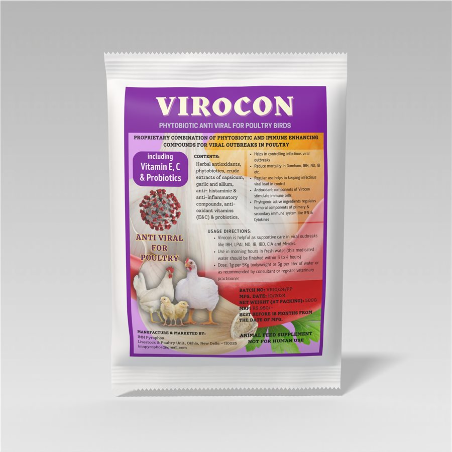
Viral outbreak support
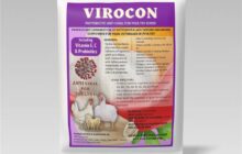
Virocon
Phytobiotic support with herbal antioxidants, capsicum, garlic and allium extracts, vitamins E and C and probiotics. Supportive care in viral outbreaks such as ND, IB, IBD, IBH, LPAI, CIA and Marek’s; helps reduce mortality.
Dose: 1 g per 5 kg body weight or 3 g per litre. Give in the morning in fresh water and let birds finish it in 3–4 hours.
500 g ₹579MRP ₹950
3. Gizzard Erosion and Ulceration Syndrome (GEU)
- Definition: A significant cause of economic losses due to gizzard lesions.
- Causative Agent: Caused by FAdV-1.
- Occurrence: Emerged in the last 20 years, affecting broilers, pullets, broiler breeders, and commercial egg-laying flocks. Can occur sporadically or in outbreaks.
- Clinical Signs: Affected birds may die with no evident clinical signs. Losses are mainly due to reduced growth rate, increased mortality, and condemnation of gizzards at slaughter.
- Gross Lesions: Gizzards may be dilated or contain brown sanguineous fluid. There are variably sized brown-black discolored areas of koilin, with erosions, ulceration, and inflammation extending to the deeper mucosa.
- Microscopic Lesions: Basophilic intranuclear inclusions typical of adenoviruses are commonly, but not always, detectable in the glandular epithelial cells. There is also thickening and vacuolation of the koilin layer due to inflammation and degeneration.
4. Egg Drop Syndrome '76 (EDS'76)
- Definition: An infectious disease of laying hens characterized by a loss of color in pigmented eggs and failure to achieve production targets, or by the production of thin-shelled, soft-shelled, or shell-less eggs in otherwise healthy-looking birds.
- Causative Agent: Caused by Duck adenovirus-1 (DAdV-1), an Atadenovirus.
- Occurrence: Widespread globally, but not historically described in North America until recently (first reported in USA in 2019).
- Clinical Signs: Primary signs are associated with egg quality issues: loss of color in pigmented eggs, thin-shelled, soft-shelled, or shell-less eggs. Egg production may drop rapidly or fail to reach expected peaks. Other mild signs can include transient dullness, watery droppings (due to oviduct fluid secretion), small eggs, watery albumen, or delayed onset of lay.
- Gross Lesions: Often minimal in natural infections. May include inactive ovaries and atrophied oviducts, though these are not consistently present. Experimentally, edema and swelling of uterine mucosal folds and exudate in the shell gland lumen have been described.
- Microscopic Lesions: In experimentally infected hens, oviduct changes include proprial edema, infiltration of inflammatory cells, atrophy of tubular glands, and degeneration/desquamation and attenuation of uterine epithelium. Intranuclear adenoviral inclusions may be seen in epithelial cells of the uterus, isthmus, and vagina. However, these acute inflammation, necrosis, or viral inclusions are rarely identified in naturally occurring outbreaks because the lesions are transient.
5. Avian Adenovirus Splenomegaly (AAS)
- Definition: A disease in chickens caused by Siadenoviruses. While details on specific chicken lesions are less emphasized, Siadenoviruses (Turkey siadenovirus A) are known to cause splenomegaly (enlarged spleen) in pheasants (Marble Spleen Disease) and turkeys (Hemorrhagic Enteritis), both of which show characteristic intranuclear inclusions in splenic cells.
Diagnosing Adenovirus Infections in Chickens
- Histopathology: The demonstration of characteristic intranuclear inclusion bodies in affected organs (e.g., liver for IBH/HPS, gizzard for GEU, oviduct for EDS) is a key diagnostic method. For IBH, HPS, and GEU, inclusions are usually readily detected.
- Virus Isolation: Isolation of the specific FAdV from affected organs (e.g., liver for IBH) can confirm the diagnosis. For EDS'76, duck/goose embryonated eggs or cell cultures are used.
- Molecular Methods (PCR): Detection of adenoviral DNA (e.g., FAdV DNA in liver for IBH) by PCR is a method of choice for direct identification and genotyping.
- Serology:
- For IBH, seroconversion to associated serotypes (FAdV-2, -8, -11) can be detected by micro-neutralization assays. However, general serological tests for group antigens are of limited value due to widespread infection and inability to differentiate pathogenic from non-pathogenic strains.
- For EDS'76, serological tests like hemagglutination inhibition (HI) are very helpful, especially in unvaccinated flocks, as infected hens typically seroconvert by the time abnormal eggs are produced.
- Electron Microscopy: Can augment routine diagnosis.
Control and Prevention in Chicken Flocks
There is no specific treatment available for avian adenovirus infections. Control strategies primarily focus on prevention:
- Biosecurity: Strict biosecurity practices are essential, including proper disinfection of barns and equipment, and good ventilation. This helps prevent horizontal spread, especially since adenoviruses are resistant to environmental factors.
- Vaccination:
- IBH/HPS: Inactivated vaccines, sometimes derived from liver homogenates of infected birds or cell cultures, are available in some countries. Live-licensed vaccines (e.g., against FAdV-8b in Australia) and autogenous vaccines (often bivalent, containing FAdV-8 and FAdV-11) are also used. Control of immunosuppressive viruses like Infectious Bursal Disease Virus (IBDV) and Chicken Anemia Virus (CAV) is also important to prevent severe IBH outbreaks, as they can enhance adenovirus pathogenicity.
- EDS'76: Inactivated vaccines, typically administered before the onset of lay, have been very successful in controlling clinical EDS. Eradication programs are also used. Minimizing contact with wild waterfowl (natural hosts) and using treated water sources are also recommended.
- Breeding Stock Management: To avoid vertical transmission, eggs from primary breeding flocks with a history of recurrent IBH in their progeny should not be used for hatching. For EDS'76, eradication has been achieved in some primary breeders.
- Environmental Resistance: Adenoviruses are relatively resistant to physical and chemical factors, making environmental persistence a challenge.
By implementing robust biosecurity measures and considering vaccination strategies where available, chicken farmers can significantly reduce the impact of these challenging adenovirus infections. IBH Infection ibh in poultry
Problem in your flock? Send photos of the birds and droppings on WhatsApp for a first opinion from Dr. Ibne Ali.
WhatsApp: 98715 84101मुर्गियों में Avian Adenovirus संक्रमण को समझें: एक पूरी गाइड
Avian adenovirus संक्रमण poultry में एक आम चिंता है, खास तौर पर दुनिया भर की मुर्गियों में। कई संक्रमण बिना लक्षण वाले (subclinical) होते हैं या किसी साफ पहचान में न आने वाले syndromes से जुड़े होते हैं, लेकिन कुछ adenoviruses अपने आप में बड़ी बीमारी पैदा करने वाले रोगाणु हैं, जो अलग पहचान वाली और आर्थिक रूप से महत्वपूर्ण बीमारियाँ करते हैं। झुंड की सेहत और उत्पादन बनाए रखने के लिए इन संक्रमणों को समझना बहुत ज़रूरी है।
मुर्गियों में Avian Adenoviruses क्या हैं?
Adenoviruses DNA viruses हैं जो संक्रमित cells के nucleus में बढ़ते हैं और वहाँ खास तरह के inclusion bodies बनाते हैं। इन पर कोई बाहरी परत (envelope) नहीं होती और इनका आकार 70 से 90 nm तक होता है। Poultry में adenoviruses को तीन genera में बाँटा गया है, और मुर्गियों को प्रभावित करने वाले मुख्य रूप से इनमें आते हैं:
- Aviadenovirus (Group I Avian adenoviruses): इस genus में Fowl adenoviruses (FAdV) आते हैं, जिनके 12 serotypes (FAdV-1 से FAdV-11) पाँच species (A-E) में बँटे हैं।
- Siadenovirus (Group II Avian adenoviruses): Turkey adenovirus A (TAdV-3), जो तीतर (pheasants) में Marble Spleen Disease करता है, मुर्गियों में Avian Adenovirus Splenomegaly भी कर सकता है।
- Atadenovirus (Group III Avian adenoviruses): Duck adenovirus-1 (DAdV-1) मुर्गियों में Egg Drop Syndrome (EDS) करता है।
मुर्गियों में संक्रमण कैसे होता है?
Adenoviruses vertical और horizontal, दोनों तरह से फैल सकते हैं।
- Vertical Transmission: संक्रमित breeders अंडों के ज़रिए virus अपने चूज़ों तक पहुँचा सकते हैं। इससे छोटे पक्षियों में outbreaks हो सकते हैं या छिपा हुआ (latent) संक्रमण रह सकता है जो बाद में, खासकर stress या peak अंडा उत्पादन के समय, फिर से सक्रिय हो जाता है।
- Horizontal Transmission: Virus बीट के साथ बाहर निकलता है, इसलिए बीट से मुँह (fecal-oral) वाला रास्ता फैलने का मुख्य तरीका है। दूषित माहौल, fomites (जैसे egg trays और transport गाड़ियाँ), काम करने वाले लोग, और सफाई के दौरान उड़ने वाली धूल भी farms के बीच या झुंड के अंदर संक्रमण फैला सकती है। कुछ adenoviruses, जैसे FAdV-4, हवा (aerosol) के ज़रिए भी फैल सकते हैं।
मुर्गियों में मुख्य Adenovirus बीमारियाँ और उनके घाव (Lesions)
Avian adenovirus संक्रमण से जुड़े घाव इस पर बहुत निर्भर करते हैं कि कौन सा virus या syndrome है। यहाँ मुर्गियों की मुख्य बीमारियों का ब्यौरा है:
1. Inclusion Body Hepatitis (IBH)
- परिभाषा: IBH छोटी उम्र की मुर्गियों की बीमारी है, जिसमें अचानक शुरुआत, बढ़ी हुई mortality, और intranuclear inclusion bodies के साथ liver में सूजन (hepatitis) होती है।
- कारण बनने वाले virus: सबसे ज़्यादा FAdV-2, FAdV-8 (8a और 8b), और FAdV-11 से होती है, लेकिन FAdV-4 (HPS के हिस्से के रूप में) और FAdV-6 से भी।
- कहाँ और कब होती है: पूरी दुनिया में पाई जाती है, आम तौर पर 2 से 5 हफ्ते के broilers में दिखती है, लेकिन इससे छोटे या बड़े पक्षियों, layers और breeders को भी हो सकती है।
- लक्षण: अक्सर पहला संकेत mortality में अचानक तेज़ बढ़ोतरी होता है (30% तक, लेकिन आम तौर पर इससे कम)। बीमार पक्षी सुस्त, बेजान, पीले/फीके (कलगी, गलकंबल, चेहरे की त्वचा) दिख सकते हैं, पंख फुलाए रहते हैं या एक जगह झुंड बनाकर बैठते हैं। पीली, चिपचिपी बीट भी दिख सकती है।
- बाहरी घाव (Gross Lesions):
- Liver: हमेशा प्रभावित होता है, सूजा हुआ, बड़ा, हल्का गुलाबी से भूरा, पीला या चितकबरा दिखता है। इसमें जगह-जगह नरम हिस्से या capsule के नीचे और अंदर petechial और ecchymotic hemorrhages (खून के छोटे-बड़े धब्बे) हो सकते हैं।
- Kidneys: अक्सर सूजी हुई, फीकी या चितकबरी।
- Bursa of Fabricius: आकार में छोटा हो सकता है।
- अन्य: हल्का hydropericardium, necrotizing pancreatitis, फीकी त्वचा, पीलापन (jaundice), टाँग और सीने की मांसपेशियों में petechial hemorrhages, खून की कमी (anemia), त्वचा/चमड़ी के नीचे की चर्बी में पीलिया (icterus), और bone marrow का खराब होना हो सकता है। Gizzard में erosions भी दिख सकते हैं।
- Microscopic घाव:
- Liver: Hepatocytes का कई जगह या बड़े हिस्से में खराब होना और मरना (necrosis)। इसकी खास पहचान hepatocytes के अंदर, खासकर necrotic हिस्सों के किनारों पर, बड़े basophilic intranuclear adenoviral inclusions का होना है।
- Kidneys: Membranous और membranoproliferative glomerulitis, कभी-कभी cortical tubules का खराब होना और necrosis।
- Bursa: Follicles का आकार छोटा और lymphoid cells की कमी।
- Pancreas: Intranuclear inclusions के साथ necrotizing pancreatitis।
- Gizzard: जगह-जगह necrotic घाव।
2. Hydropericardium/Hepatitis Syndrome (HPS)
- परिभाषा: IBH जैसी ही, लेकिन ज़्यादा तेज़ (acute) और ज़्यादा mortality वाली, जिसमें दिल के चारों ओर काफी पानी भर जाता है।
- कारण बनने वाला virus: खतरनाक FAdV-4 strains से होती है।
- कहाँ और कब होती है: मुख्य रूप से 3 से 6 हफ्ते के broiler चूज़ों में, लेकिन layers और breeders में भी रिपोर्ट हुई है।
- लक्षण: तेज़ी से बढ़ती mortality (20-80%), सुस्ती, उदासी, पंख फुलाना, झुंड बनाकर बैठना, पेट के बल लेटना, और बाद की stage में आँखें बंद रहना।
- बाहरी घाव (Gross Lesions): सबसे खास बात है दिल की थैली (pericardial sac) में 10 ml तक पानी जैसा द्रव भर जाना (hydropericardium)। Liver का रंग बदला हुआ और बड़ा होता है, और उसमें जगह-जगह necrosis और खून के धब्बे हो सकते हैं। फेफड़ों में सूजन (edema) और खून जमाव हो सकता है, और urate जमने से kidneys फीकी और tubules बढ़े हुए हो सकते हैं।
- Microscopic घाव: IBH की तरह ही hepatocytes में intranuclear inclusion bodies के साथ गंभीर necrotizing hepatitis। साथ ही, virus lymphoid tissues को निशाना बनाता है, जिससे bursa, thymus और spleen में lymphoid अंगों का खराब होना और lymphocytes की कमी (lymphocytolysis) होती है।
वायरल बीमारी में सहारा
Virocon
हर्बल एंटीऑक्सीडेंट, कैप्सिकम, लहसुन और एलियम एक्सट्रैक्ट, विटामिन E और C और प्रोबायोटिक वाला फाइटोबायोटिक। ND, IB, IBD, IBH, LPAI, CIA और मैरेक्स जैसी वायरल बीमारियों में सहायक इलाज; मृत्यु घटाने में मदद करता है।
मात्रा: 1 ग्राम प्रति 5 किलो वज़न या 3 ग्राम प्रति लीटर। सुबह ताज़े पानी में दें और 3–4 घंटे में खत्म होने दें।
500 g ₹579MRP ₹950
3. Gizzard Erosion and Ulceration Syndrome (GEU)
- परिभाषा: Gizzard के घावों की वजह से आर्थिक नुकसान का एक बड़ा कारण।
- कारण बनने वाला virus: FAdV-1 से होता है।
- कहाँ और कब होता है: पिछले 20 सालों में सामने आया है, broilers, pullets, broiler breeders और commercial अंडा देने वाले झुंडों को प्रभावित करता है। यह कभी-कभार (sporadic) या outbreaks के रूप में हो सकता है।
- लक्षण: बीमार पक्षी बिना किसी साफ लक्षण के मर सकते हैं। नुकसान मुख्य रूप से बढ़वार की कम दर, बढ़ी mortality, और कटाई (slaughter) के समय gizzards के रिजेक्ट होने से होता है।
- बाहरी घाव (Gross Lesions): Gizzard फैला हुआ हो सकता है या उसमें भूरा खूनी द्रव हो सकता है। Koilin (gizzard की अंदरूनी परत) पर अलग-अलग आकार के भूरे-काले रंग के हिस्से, erosions, ulcers और सूजन होती है जो गहरी mucosa तक जाती है।
- Microscopic घाव: Glandular epithelial cells में adenoviruses वाले basophilic intranuclear inclusions अक्सर, लेकिन हमेशा नहीं, दिखते हैं। सूजन और खराबी के कारण koilin परत मोटी और खोखली (vacuolation) भी हो जाती है।
4. Egg Drop Syndrome '76 (EDS'76)
- परिभाषा: अंडा देने वाली मुर्गियों की एक संक्रामक बीमारी, जिसमें रंगीन अंडों का रंग उड़ जाता है और उत्पादन का लक्ष्य पूरा नहीं होता, या बाकी तौर पर स्वस्थ दिखने वाले पक्षी पतले छिलके वाले, नरम छिलके वाले या बिना छिलके के अंडे देते हैं।
- कारण बनने वाला virus: Duck adenovirus-1 (DAdV-1) से होती है, जो एक Atadenovirus है।
- कहाँ और कब होती है: पूरी दुनिया में फैली है, लेकिन North America में हाल तक इसका वर्णन नहीं था (USA में पहली बार 2019 में रिपोर्ट हुई)।
- लक्षण: मुख्य लक्षण अंडे की quality की समस्याओं से जुड़े हैं: रंगीन अंडों का रंग उड़ना, पतले छिलके वाले, नरम छिलके वाले या बिना छिलके के अंडे। अंडा उत्पादन तेज़ी से गिर सकता है या उम्मीद के peak तक नहीं पहुँचता। दूसरे हल्के लक्षणों में थोड़े समय की सुस्ती, पतली बीट (oviduct से द्रव निकलने के कारण), छोटे अंडे, पानी जैसी सफेदी (albumen), या अंडा देना देर से शुरू होना शामिल हो सकते हैं।
- बाहरी घाव (Gross Lesions): प्राकृतिक संक्रमण में अक्सर बहुत कम होते हैं। इसमें निष्क्रिय अंडाशय (ovaries) और सिकुड़ी हुई oviducts हो सकती हैं, हालाँकि ये हमेशा नहीं मिलतीं। प्रयोगों में uterus की mucosal परतों में सूजन (edema) और shell gland के अंदर स्राव (exudate) देखा गया है।
- Microscopic घाव: प्रयोग में संक्रमित मुर्गियों की oviduct में proprial edema, सूजन वाली cells का जमाव, tubular glands का सिकुड़ना, और uterus की epithelium का खराब होना/झड़ना और पतला होना देखा गया है। Uterus, isthmus और vagina की epithelial cells में intranuclear adenoviral inclusions दिख सकते हैं। लेकिन ये तेज़ सूजन, necrosis या viral inclusions प्राकृतिक outbreaks में बहुत कम पहचाने जाते हैं क्योंकि ये घाव थोड़े समय के लिए ही रहते हैं।
5. Avian Adenovirus Splenomegaly (AAS)
- परिभाषा: मुर्गियों में Siadenoviruses से होने वाली बीमारी। मुर्गियों के खास घावों पर ज़्यादा जानकारी नहीं दी जाती, लेकिन Siadenoviruses (Turkey siadenovirus A) तीतर (Marble Spleen Disease) और टर्की (Hemorrhagic Enteritis) में splenomegaly (बढ़ी हुई spleen) करने के लिए जाने जाते हैं, और दोनों में spleen की cells में खास intranuclear inclusions दिखते हैं।
मुर्गियों में Adenovirus संक्रमण की जाँच
- Histopathology: प्रभावित अंगों में खास intranuclear inclusion bodies दिखाना (जैसे IBH/HPS के लिए liver, GEU के लिए gizzard, EDS के लिए oviduct) जाँच का मुख्य तरीका है। IBH, HPS और GEU में inclusions आम तौर पर आसानी से दिख जाते हैं।
- Virus Isolation: प्रभावित अंगों (जैसे IBH के लिए liver) से खास FAdV को अलग करके जाँच की पुष्टि हो सकती है। EDS'76 के लिए बत्तख/हंस के embryonated eggs या cell cultures इस्तेमाल होते हैं।
- Molecular तरीके (PCR): PCR से adenoviral DNA की पहचान (जैसे IBH के लिए liver में FAdV DNA) सीधी पहचान और genotyping के लिए सबसे पसंदीदा तरीका है।
- Serology:
- IBH के लिए, संबंधित serotypes (FAdV-2, -8, -11) के खिलाफ seroconversion micro-neutralization assays से पता लगाया जा सकता है। लेकिन group antigens के लिए आम serological tests का फायदा सीमित है, क्योंकि संक्रमण बहुत फैला हुआ है और ये बीमारी करने वाले और न करने वाले strains में फर्क नहीं कर पाते।
- EDS'76 के लिए hemagglutination inhibition (HI) जैसे serological tests बहुत मददगार हैं, खासकर बिना vaccination वाले झुंडों में, क्योंकि जब तक असामान्य अंडे आने लगते हैं, तब तक संक्रमित मुर्गियों में आम तौर पर seroconversion हो चुका होता है।
- Electron Microscopy: नियमित जाँच में मदद कर सकती है।
मुर्गियों के झुंड में नियंत्रण और बचाव
Avian adenovirus संक्रमण का कोई खास इलाज उपलब्ध नहीं है। नियंत्रण की रणनीतियाँ मुख्य रूप से बचाव पर टिकी हैं:
- Biosecurity: सख्त biosecurity बहुत ज़रूरी है, जिसमें shed और उपकरणों का सही disinfection और अच्छा ventilation शामिल है। इससे horizontal फैलाव रुकता है, खासकर इसलिए क्योंकि adenoviruses माहौल के असर को झेल लेते हैं।
- Vaccination:
- IBH/HPS: कुछ देशों में inactivated vaccines उपलब्ध हैं, जो कभी-कभी संक्रमित पक्षियों के liver homogenate या cell cultures से बनती हैं। Live-licensed vaccines (जैसे Australia में FAdV-8b के खिलाफ) और autogenous vaccines (अक्सर bivalent, जिनमें FAdV-8 और FAdV-11 होते हैं) भी इस्तेमाल होती हैं। गंभीर IBH outbreaks रोकने के लिए Infectious Bursal Disease Virus (IBDV) और Chicken Anemia Virus (CAV) जैसे immunity दबाने वाले viruses का नियंत्रण भी ज़रूरी है, क्योंकि ये adenovirus की बीमारी करने की ताकत बढ़ा सकते हैं।
- EDS'76: Inactivated vaccines, जो आम तौर पर अंडा शुरू होने से पहले दी जाती हैं, clinical EDS को नियंत्रित करने में बहुत सफल रही हैं। इसे खत्म करने के कार्यक्रम (eradication programs) भी चलाए जाते हैं। जंगली जलपक्षियों (प्राकृतिक host) से संपर्क कम करने और treated पानी के स्रोत इस्तेमाल करने की भी सलाह दी जाती है।
- Breeding Stock का प्रबंधन: Vertical transmission से बचने के लिए, जिन primary breeding झुंडों के चूज़ों में बार-बार IBH होने का इतिहास है, उनके अंडे hatching के लिए इस्तेमाल नहीं करने चाहिए। EDS'76 के लिए कुछ primary breeders में इसे खत्म किया जा चुका है।
- माहौल में टिके रहने की क्षमता: Adenoviruses भौतिक और रासायनिक कारकों के प्रति काफी मज़बूत होते हैं, इसलिए माहौल में इनका बने रहना एक चुनौती है।
मज़बूत biosecurity उपाय अपनाकर और जहाँ उपलब्ध हो वहाँ vaccination रणनीतियों पर ध्यान देकर, मुर्गी पालक इन मुश्किल adenovirus संक्रमणों के असर को काफी कम कर सकते हैं। IBH Infection, poultry में ibh
फ़ार्म में कोई दिक्कत? पक्षियों और बीट की फ़ोटो WhatsApp पर भेजिए, डॉ. इब्ने अली से पहली राय लीजिए।
WhatsApp: 98715 84101Murgiyon me Avian Adenovirus Infection ko Samjhein: Ek Poori Guide
Avian adenovirus infection poultry me ek common chinta hai, khaas kar duniya bhar ki murgiyon me. Kai infection bina lakshan wale (subclinical) hote hain ya kisi saaf pehchan me na aane wale syndromes se jude hote hain, lekin kuch adenoviruses khud me bade pathogen hain jo alag pehchan wali aur aarthik roop se important bimariyan karte hain. Flock ki health aur production banaye rakhne ke liye in infections ko samajhna bahut zaroori hai.
Murgiyon me Avian Adenoviruses kya hain?
Adenoviruses DNA viruses hain jo infected cells ke nucleus me badhte hain aur wahan khaas tarah ke inclusion bodies banate hain. In par koi bahari parat (envelope) nahi hoti aur inka size 70 se 90 nm tak hota hai. Poultry me adenoviruses ko teen genera me baanta gaya hai, aur murgiyon ko affect karne wale mainly inme aate hain:
- Aviadenovirus (Group I Avian adenoviruses): Is genus me Fowl adenoviruses (FAdV) aate hain, jinke 12 serotypes (FAdV-1 se FAdV-11) paanch species (A-E) me baante gaye hain.
- Siadenovirus (Group II Avian adenoviruses): Turkey adenovirus A (TAdV-3), jo teetar (pheasants) me Marble Spleen Disease karta hai, murgiyon me Avian Adenovirus Splenomegaly bhi kar sakta hai.
- Atadenovirus (Group III Avian adenoviruses): Duck adenovirus-1 (DAdV-1) murgiyon me Egg Drop Syndrome (EDS) karta hai.
Murgiyon me Infection kaise hota hai?
Adenoviruses vertical aur horizontal, dono tarah se fail sakte hain.
- Vertical Transmission: Infected breeders ando ke zariye virus apne chuzon tak pahuncha sakte hain. Isse chhote birds me outbreak ho sakta hai ya chhupa hua (latent) infection reh sakta hai jo baad me, khaas kar stress ya peak egg production ke time, phir se active ho jata hai.
- Horizontal Transmission: Virus beet ke saath bahar nikalta hai, isliye beet se munh (fecal-oral) wala rasta failne ka main tareeka hai. Contaminated mahaul, fomites (jaise egg trays aur transport gaadiyan), kaam karne wale log, aur safai ke time udne wali dhool bhi farms ke beech ya flock ke andar infection faila sakti hai. Kuch adenoviruses, jaise FAdV-4, hawa (aerosol) ke zariye bhi fail sakte hain.
Murgiyon me Mukhya Adenovirus Bimariyan aur unke Lesions
Avian adenovirus infection se jude lesions is baat par bahut depend karte hain ki kaun sa virus ya syndrome hai. Yahan murgiyon ki main bimariyon ka breakdown hai:
1. Inclusion Body Hepatitis (IBH)
- Definition: IBH chhoti age ki murgiyon ki bimari hai jisme achanak shuruat, badhi hui mortality, aur intranuclear inclusion bodies ke saath liver me sujan (hepatitis) hoti hai.
- Karan banne wale virus: Sabse zyada FAdV-2, FAdV-8 (8a aur 8b), aur FAdV-11 se hoti hai, lekin FAdV-4 (HPS ke hisse ke roop me) aur FAdV-6 se bhi.
- Kahan aur kab hoti hai: Poori duniya me milti hai, aam taur par 2 se 5 hafte ke broilers me dikhti hai, lekin isse chhote ya bade birds, layers aur breeders ko bhi ho sakti hai.
- Lakshan: Aksar pehla sign mortality me achanak tez badhotri hota hai (30% tak, lekin aam taur par isse kam). Bimar birds sust, bejaan, peele/feeke (kalgi, wattles, chehre ki skin) dikh sakte hain, pankh phulaye rehte hain ya ek jagah jhund bana kar baithte hain. Peeli, chipchipi beet bhi dikh sakti hai.
- Gross Lesions:
- Liver: Hamesha affect hota hai, suja hua, bada, halka gulabi se bhoora, peela ya chitkabra dikhta hai. Isme jagah-jagah naram hisse ya capsule ke neeche aur andar petechial aur ecchymotic hemorrhages (khoon ke chhote-bade dhabbe) ho sakte hain.
- Kidneys: Aksar suji hui, feeki ya chitkabri.
- Bursa of Fabricius: Size me chhota ho sakta hai.
- Anya: Halka hydropericardium, necrotizing pancreatitis, feeki skin, peelapan (jaundice), taang aur breast ki muscles me petechial hemorrhages, khoon ki kami (anemia), skin/chamdi ke neeche ki charbi me peeliya (icterus), aur bone marrow ka kharab hona ho sakta hai. Gizzard me erosions bhi dikh sakte hain.
- Microscopic Lesions:
- Liver: Hepatocytes ka kai jagah ya bade hisse me kharab hona aur marna (necrosis). Iski khaas pehchan hepatocytes ke andar, khaas kar necrotic hisson ke kinaron par, bade basophilic intranuclear adenoviral inclusions ka hona hai.
- Kidneys: Membranous aur membranoproliferative glomerulitis, kabhi-kabhi cortical tubules ka kharab hona aur necrosis.
- Bursa: Follicles ka size chhota aur lymphoid cells ki kami.
- Pancreas: Intranuclear inclusions ke saath necrotizing pancreatitis.
- Gizzard: Jagah-jagah necrotic lesions.
2. Hydropericardium/Hepatitis Syndrome (HPS)
- Definition: IBH jaisi hi, lekin zyada tez (acute) aur zyada mortality wali, jisme dil ke chaaron taraf kaafi paani bhar jata hai.
- Karan banne wala virus: Khatarnak FAdV-4 strains se hoti hai.
- Kahan aur kab hoti hai: Mainly 3 se 6 hafte ke broiler chuzon me, lekin layers aur breeders me bhi report hui hai.
- Lakshan: Tezi se badhti mortality (20-80%), susti, udaasi, pankh phulana, jhund bana kar baithna, pet ke bal letna, aur baad ki stage me aankhein band rehna.
- Gross Lesions: Sabse khaas baat hai dil ki thaili (pericardial sac) me 10 ml tak paani jaisa fluid bhar jana (hydropericardium). Liver ka rang badla hua aur bada hota hai, aur usme jagah-jagah necrosis aur khoon ke dhabbe ho sakte hain. Lungs me sujan (edema) aur congestion ho sakta hai, aur urate jamne se kidneys feeki aur tubules bade hue ho sakte hain.
- Microscopic Lesions: IBH ki tarah hi hepatocytes me intranuclear inclusion bodies ke saath serious necrotizing hepatitis. Saath hi, virus lymphoid tissues ko target karta hai, jisse bursa, thymus aur spleen me lymphoid organs ka kharab hona aur lymphocytes ki kami (lymphocytolysis) hoti hai.
Viral bimari me sahara
Virocon
Herbal antioxidant, capsicum, lehsun aur allium extract, vitamin E aur C aur probiotic wala phytobiotic. ND, IB, IBD, IBH, LPAI, CIA aur Marek’s jaisi viral bimariyon me sahayak ilaaj; maut ghatane me madad karta hai.
Matra: 1 g prati 5 kg wazan ya 3 g prati litre. Subah taaze paani me dijiye aur 3–4 ghante me khatam hone dijiye.
500 g ₹579MRP ₹950
3. Gizzard Erosion and Ulceration Syndrome (GEU)
- Definition: Gizzard ke lesions ki wajah se aarthik nuksan ka ek bada karan.
- Karan banne wala virus: FAdV-1 se hota hai.
- Kahan aur kab hota hai: Pichhle 20 saalon me saamne aaya hai, broilers, pullets, broiler breeders aur commercial egg laying flocks ko affect karta hai. Ye kabhi-kabhaar (sporadic) ya outbreak ke roop me ho sakta hai.
- Lakshan: Bimar birds bina kisi saaf lakshan ke mar sakte hain. Nuksan mainly growth rate kam hone, badhi mortality, aur slaughter ke time gizzards reject hone se hota hai.
- Gross Lesions: Gizzard faila hua ho sakta hai ya usme bhoora khooni fluid ho sakta hai. Koilin (gizzard ki andar ki parat) par alag-alag size ke bhoore-kaale rang ke hisse, erosions, ulcers aur sujan hoti hai jo gehri mucosa tak jaati hai.
- Microscopic Lesions: Glandular epithelial cells me adenoviruses wale basophilic intranuclear inclusions aksar, lekin hamesha nahi, dikhte hain. Sujan aur kharabi ki wajah se koilin parat moti aur khokhli (vacuolation) bhi ho jaati hai.
4. Egg Drop Syndrome '76 (EDS'76)
- Definition: Egg dene wali murgiyon ki ek infectious bimari, jisme rangeen ando ka rang ud jata hai aur production target poora nahi hota, ya baaki tarah se healthy dikhne wale birds patle chhilke wale, naram chhilke wale ya bina chhilke ke ande dete hain.
- Karan banne wala virus: Duck adenovirus-1 (DAdV-1) se hoti hai, jo ek Atadenovirus hai.
- Kahan aur kab hoti hai: Poori duniya me faili hai, lekin North America me haal tak iska zikr nahi tha (USA me pehli baar 2019 me report hui).
- Lakshan: Main lakshan egg quality ki problems se jude hain: rangeen ando ka rang udna, patle chhilke wale, naram chhilke wale ya bina chhilke ke ande. Egg production tezi se gir sakta hai ya expected peak tak nahi pahunchta. Doosre halke lakshan me thode samay ki susti, patli beet (oviduct se fluid nikalne ki wajah se), chhote ande, paani jaisi safedi (albumen), ya laying der se shuru hona shamil ho sakte hain.
- Gross Lesions: Natural infection me aksar bahut kam hote hain. Isme inactive ovaries aur sikudi hui oviducts ho sakti hain, haalanki ye hamesha nahi miltin. Experiments me uterus ki mucosal parton me sujan (edema) aur shell gland ke andar exudate dekha gaya hai.
- Microscopic Lesions: Experiment me infected murgiyon ki oviduct me proprial edema, sujan wali cells ka jamav, tubular glands ka sikudna, aur uterus ki epithelium ka kharab hona/jhadna aur patla hona dekha gaya hai. Uterus, isthmus aur vagina ki epithelial cells me intranuclear adenoviral inclusions dikh sakte hain. Lekin ye tez sujan, necrosis ya viral inclusions natural outbreaks me bahut kam pehchane jaate hain kyunki ye lesions thode samay ke liye hi rehte hain.
5. Avian Adenovirus Splenomegaly (AAS)
- Definition: Murgiyon me Siadenoviruses se hone wali bimari. Murgiyon ke khaas lesions par zyada jaankari nahi di jaati, lekin Siadenoviruses (Turkey siadenovirus A) teetar (Marble Spleen Disease) aur turkey (Hemorrhagic Enteritis) me splenomegaly (badhi hui spleen) karne ke liye jaane jaate hain, aur dono me spleen ki cells me khaas intranuclear inclusions dikhte hain.
Murgiyon me Adenovirus Infection ka Diagnosis
- Histopathology: Affected organs me khaas intranuclear inclusion bodies dikhana (jaise IBH/HPS ke liye liver, GEU ke liye gizzard, EDS ke liye oviduct) diagnosis ka main tareeka hai. IBH, HPS aur GEU me inclusions aam taur par aasani se dikh jaate hain.
- Virus Isolation: Affected organs (jaise IBH ke liye liver) se specific FAdV ko alag karke diagnosis confirm ho sakta hai. EDS'76 ke liye batakh/hans ke embryonated eggs ya cell cultures use hote hain.
- Molecular Methods (PCR): PCR se adenoviral DNA ki pehchan (jaise IBH ke liye liver me FAdV DNA) seedhi pehchan aur genotyping ke liye sabse pasandida tareeka hai.
- Serology:
- IBH ke liye, related serotypes (FAdV-2, -8, -11) ke khilaf seroconversion micro-neutralization assays se pata lagaya ja sakta hai. Lekin group antigens ke liye aam serological tests ka fayda limited hai, kyunki infection bahut faila hua hai aur ye bimari karne wale aur na karne wale strains me fark nahi kar paate.
- EDS'76 ke liye hemagglutination inhibition (HI) jaise serological tests bahut madadgaar hain, khaas kar bina vaccination wale flocks me, kyunki jab tak abnormal ande aane lagte hain, tab tak infected murgiyon me aam taur par seroconversion ho chuka hota hai.
- Electron Microscopy: Routine diagnosis me madad kar sakti hai.
Murgiyon ke Flock me Control aur Bachav
Avian adenovirus infection ka koi specific ilaaj available nahi hai. Control strategies mainly bachav par tiki hain:
- Biosecurity: Sakht biosecurity bahut zaroori hai, jisme shed aur equipment ka sahi disinfection aur achha ventilation shamil hai. Isse horizontal failav rukta hai, khaas kar isliye kyunki adenoviruses mahaul ke asar ko jhel lete hain.
- Vaccination:
- IBH/HPS: Kuch deshon me inactivated vaccines available hain, jo kabhi-kabhi infected birds ke liver homogenate ya cell cultures se banti hain. Live-licensed vaccines (jaise Australia me FAdV-8b ke khilaf) aur autogenous vaccines (aksar bivalent, jinme FAdV-8 aur FAdV-11 hote hain) bhi use hoti hain. Serious IBH outbreaks rokne ke liye Infectious Bursal Disease Virus (IBDV) aur Chicken Anemia Virus (CAV) jaise immunity dabane wale viruses ka control bhi zaroori hai, kyunki ye adenovirus ki bimari karne ki taakat badha sakte hain.
- EDS'76: Inactivated vaccines, jo aam taur par laying shuru hone se pehle di jaati hain, clinical EDS ko control karne me bahut safal rahi hain. Ise khatam karne ke programs (eradication programs) bhi chalaye jaate hain. Jungli paani wale pakshiyon (natural host) se contact kam karne aur treated paani ke source use karne ki bhi salah di jaati hai.
- Breeding Stock Management: Vertical transmission se bachne ke liye, jin primary breeding flocks ke chuzon me baar-baar IBH hone ka itihaas hai, unke ande hatching ke liye use nahi karne chahiye. EDS'76 ke liye kuch primary breeders me ise khatam kiya ja chuka hai.
- Mahaul me tike rehne ki kshamta: Adenoviruses physical aur chemical factors ke saamne kaafi mazboot hote hain, isliye mahaul me inka bane rehna ek challenge hai.
Mazboot biosecurity upay apna kar aur jahan available ho wahan vaccination strategies par dhyan dekar, murgi paalak in mushkil adenovirus infections ke asar ko kaafi kam kar sakte hain. IBH Infection, poultry me ibh
Farm me koi dikkat? Pakshiyon aur beet ki photo WhatsApp par bhejiye, Dr. Ibne Ali se pehli raay lijiye.
WhatsApp: 98715 84101